Small lipid droplets are rigid enough to indent a nucleus, dilute the lamina, and cause rupture

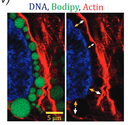
The nucleus in many cell types is a stiff organelle, but fat-filled lipid droplets (FDs) in cytoplasm are seen to indent and displace the nucleus. FDs are phase-separated liquids with a poorly understood interfacial tension γ that determines how FDs interact with other organelles. Here, micron-sized FDs remain spherical as they indent peri-nuclear actomyosin and the nucleus, while causing local dilution of Lamin-B1 independent of Lamin-A,C and sometimes triggering nuclear rupture. Focal accumulation of the cytosolic DNA sensor cGAS at the rupture site is accompanied by sustained mislocalization of DNA repair factors to cytoplasm, increased DNA damage, and delayed cell cycle. Macrophages show FDs and engulfed rigid beads cause similar indentation dilution. Spherical shapes of small FDs indicate a high γ, which we measure for FDs mechanically isolated from fresh adipose tissue as ∼40 mN/m. This value is far higher than that of protein condensates, but typical of oils in water and sufficiently rigid to perturb cell structures including nuclei.

Tension-suppressed degradation of collagen controls tissue stiffness scaling with fibrillar collagen

Extremely soft tissues such as developing hearts or adult brain contain far less collagen than highly stiff adult tissues such as tendons, but cell and molecular mechanisms for such homeostatic differences remain unclear. We hypothesized that cell-generated or exogenous forces combine with tension-suppressed collagen degradation in order to sculpt extracellular matrix (ECM) collagen levels in tissues. For various mature mice tissues and beating embryonic chick hearts, we find collagen-sensitive second harmonic generation (SHG) image intensity scales non-linearly versus tissue stiffness, aligning well with the results from cellularized gels of collagen. Chick hearts beating at ∼5% strain maintain collagen levels until their contractile strain is suppressed by myosin-II inhibition and endogenous matrix metalloproteinases (MMPs) then degrade collagens within ∼30-60 minutes – based on SHG and mass spectrometry proteomics. Although tendons composed of oriented collagen fibrils exhibit heterogeneous strain distributions upon deformation, the addition of exogenous MMP or bacterial collagenase suppresses collagen degradation for strains within physiological limits (i.e., up to ∼5-8%). Sequestration of collagen cleavage sites by tissue strain is a likely mechanism because molecular permeation and mobility prove strain-independent whereas artificial collagen cross-links accelerate strain-dependent collagen degradation via collagen molecular unfolding. Tension-suppressed degradation of collagen thus underlies tissue stiffness scaling.

Matrix Elasticity Directs Stem Cell Lineage Specification

Microenvironments appear important in stem cell lineage specification but can be difficult to adequately characterize or control with soft tissues. Naive mesenchymal stem cells (MSCs) are shown here to specify lineage and commit to phenotypes with extreme sensitivity to tissue-level elasticity. Soft matrices that mimic brain are neurogenic, stiffer matrices that mimic muscle are myogenic, and comparatively rigid matrices that mimic collagenous bone prove osteogenic. During the initial week in culture, reprogramming of these lineages is possible with addition of soluble induction factors, but after several weeks in culture, the cells commit to the lineage specified by matrix elasticity, consistent with the elasticity-insensitive commitment of differentiated cell types. Inhibition of nonmuscle myosin II blocks all elasticity-directed lineage specification–without strongly perturbing many other aspects of cell function and shape. The results have significant implications for understanding physical effects of the in vivo microenvironment and also for therapeutic uses of stem cells.